
This case study explores how leading hospitals, diagnostic pathology labs, and research institutions leverage the Pannoramic 250 Flash III DX, Pannoramic 150 DX, Pannoramic 75 DX, Pannoramic MIDI III DX, and Pannoramic Flash Desk DX to overcome IHC challenges, optimize workflows, and enhance diagnostic confidence.
Key Challenges in Diagnostic Immunohistochemistry
Inconsistent Staining Interpretation & Reproducibility Issues
• Variability in staining intensity, background noise, and antigen retrieval processes can impact results.
• Manual interpretation introduces inter-reader variability, affecting diagnostic consistency.
High Slide Volume & Processing Bottlenecks
• IHC laboratories handle hundreds of slides daily, requiring high-throughput imaging solutions.
• Traditional microscopy slows workflow efficiency and increases turnaround times.
High-Resolution Imaging for Biomarker Quantification
• Detecting subtle protein expression levels requires ultra-resolution whole slide imaging.
• Low-quality images can compromise clinical decisions and biomarker research.
Integration with LIS/HIS & Remote Consultations
• Efficient data management and seamless LIS/HIS connectivity are essential for pathology workflows.
• Digital IHC requires secure, remote-access telepathology tools for second opinions and expert collaboration.
Optimizing Clinical Pathology
Solutions
3DHISTECH’s Pannoramic DX Whole Slide Imaging Solutions deliver:
• High-resolution IHC scanning with brightfield, AI-enhanced imaging, and Z-stack capabilities
• Automated tissue detection for standardized, high-quality IHC image acquisition
• High-throughput processing of up to 300 slides per batch, minimizing delays
• DICOM-compatible format for LIS/HIS connectivity and regulatory compliance
• Telepathology integration for remote IHC diagnostics and collaboration
1. Pannoramic 250 Flash III DX – High-Speed, High-Resolution IHC Imaging
Best for: Large hospitals and high-throughput IHC labs requiring batch processing.
• Supports up to 300 slides per batch for efficient high-throughput IHC workflows.
• AI-driven tissue detection and automated batch scanning enhance reproducibility.
• Brightfield scanning at 0.1 um/pixel resolution captures detailed biomarker expression.
• Z-stack imaging for precise depth visualization, crucial for antigen localization.
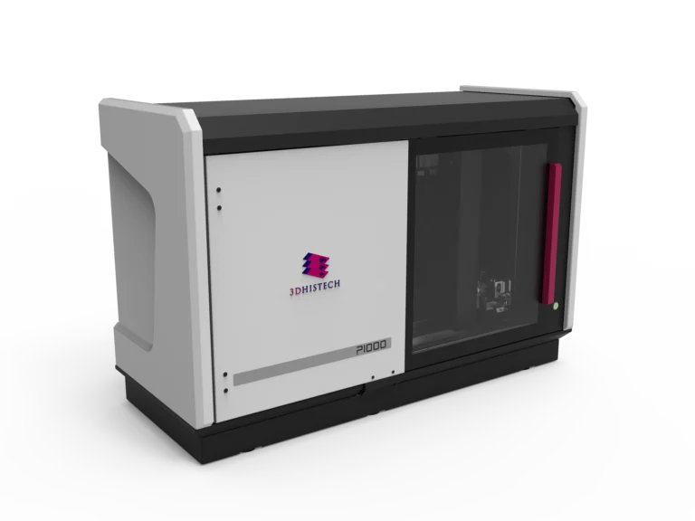
Pannoramic 1000 DX Digital Scanner
2. Pannoramic 150 DX – Precision IHC Imaging with High-Resolution Analysis
Best for: Mid-sized pathology labs focused on standardizing IHC biomarker quantification.
• 150-slide automated capacity for optimized workflow efficiency.
• 0.1 um/pixel ultra-high resolution at 100x magnification for biomarker clarity.
• Multilayer (Z-stack) and extended focus scanning improve antigen visualization.
• Automated AI-powered tissue detection minimizes operator-dependent variability.
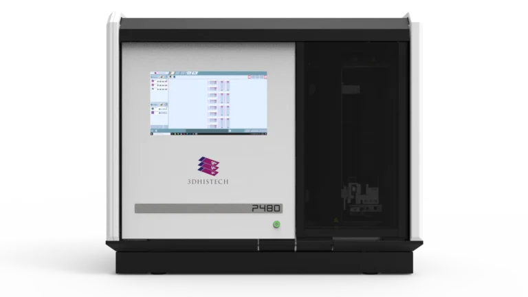
Pannoramic 480 DX Digital Scanner
3. Pannoramic 75 DX – Compact, High-Throughput IHC Solution
Best for: Medium-sized labs needing ultra-resolution IHC scanning in a space-efficient design.
• Scans up to 75 slides per batch, ideal for mid-sized IHC workflows.
• I 0.1 um/pixel resolution at 100x magnification captures fine cellular details.
• Automated Z-stack scanning ensures optimal antigen-antibody visualization.
• AI-powered slide detection and DICOM compatibility streamline workflow integration.
Pannoramic 250 Flash III DX Digital Scanner
4. Pannoramic MIDI III DX – High-Speed IHC Imaging with Flash Illumination
Best for: Labs needing high-speed scanning with brightfield clarity.
• 12-slide capacity with ultra-fast brightfield scanning at 0.12 um/pixel.
• Flash illumination technology ensures high-contrast, well-defined IHC images.
• AI-driven tissue recognition and automated focus layering improve biomarker accuracy.
• Seamless LIS/HIS integration and DICOM compatibility.
Pannoramic 250 Flash III DX Digital Scanner
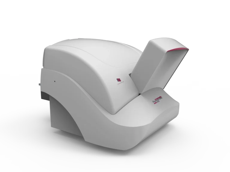
5. Pannoramic Flash Desk DX – Specialized IHC Scanner for Rapid Diagnostics
Best for: Intraoperative consultations and small-scale IHC research applications.
• Single-slide, high-resolution scanning for immediate IHC assessment.
• Flash illumination ensures fast, high-quality brightfield imaging.
• 0.12 um/pixel ultra-resolution at 82x magnification for detailed biomarker analysis.
• Compact, space-saving design ideal for intraoperative and research pathology settings.
• Intraoperative IHC diagnostics – Quick assessment of biopsy margins and tumor markers during surgery.
• Custom-stained IHC slides – Ideal for research labs analyzing experimental biomarkers.
• Point-of-care pathology – Small, low-throughput labs needing immediate, highresolution IHC analysis.
Pannoramic 250 Flash III DX Digital Scanner
Optimizing Clinical Pathology
Key Benefits for IHC Pathology Labs
Standardized, High-Quality IHC Imaging
• AI-powered automated tissue detection ensures consistent staining interpretation.
• Ultra-resolution brightfield scanning captures antigen-antibody interactions precisely.
Faster, More Reliable IHC Diagnostics
• High-throughput capacity reduces turnaround times for clinical and research applications. • Batch processing and automated workflows eliminate bottlenecks in IHC imaging.
Advanced Image Analysis for Biomarker Quantification
• 0.1 um/pixel ultra-resolution scanning allows precise IHC marker assessment.
• Z-stack imaging provides depth-focused antigen localization in tissue sections.
Seamless LIS/HIS Integration for Digital Pathology Workflows
• DICOM format support ensures easy data transfer and regulatory compliance.
• Telepathology capabilities enable remote expert collaboration and second opinions.
Cost-Effective, Scalable Solutions for Any Lab Size
• From single-slide analysis to 300-slide batch processing, solutions scale with lab demand.
• Compact and high-capacity scanners meet varied diagnostic pathology requirements.
Pannoramic DX Large Volume Scanner for Diagnostic Immunohistochemistry
For high-volume IHC laboratories, selecting the right high-capacity whole slide imaging scanner is essential to ensure efficient, precise, and reproducible biomarker analysis. The Pannoramic 480 DX Digital Scanner provides automated, AI-driven high-throughput scanning, making it an optimal solution for large-scale immunohistochemistry workflows.
Pannoramic 480 DX Digital Scanner – High-Speed IHC Imaging at Scale
Best for: Large hospitals, high-throughput IHC pathology labs, and biomarker centers needing automated, batch processing.
Key Features for IHC Applications
- Supports up to 480 slides per batch – Ideal for large-scale IHC workflows.
- Brightfield and polarization imaging – Ensures accurate biomarker expression visualization.
- Ultra-resolution imaging (40x / 80x, 50x / 100x magnification) – Detects subtle protein expression changes in IHC-stained tissue.
- Z-stack imaging for multilayer visualization – Enhances antigen depth and cellular localization analysis.
- AI-powered tissue detection – Standardizes scanning accuracy and minimizes human variability.
- Integrated touch display – Provides real-time quality control and rapid first-level IHC review.
- Precision anti-vibration base – Ensures stable, high-quality imaging for consistent staining interpretation.
- Seamless LIS/HIS integration and DICOM compatibility – Fully integrates with clinical workflows.
A Digital Pathology Transformation for IHC Diagnostics
By adopting 3DHISTECH’s Pannoramic DX Whole Slide Imaging Solutions, diagnostic IHC labs, hospitals, and research institutions have significantly improved workflow efficiency, biomarker quantification, and diagnostic standardization.
With automated scanning, ultra-resolution imaging, and seamless LIS/HIS integration, these scanners eliminate IHC workflow inefficiencies, enhance biomarker visualization, and support remote digital pathology collaborations.