
To address these challenges, 3DHISTECH’s digital pathology solutions, including high-performance digital pathology scanners, TMA technology, and AI-powered software, provide scalable, high-resolution, and automated workflows to enhance toxicology research, improve efficiency, and accelerate discovery.
Key Challenges in Toxicology and Environmental Science Research
Limited Throughput and Reproducibility
Toxicology research often involves large-scale screening of tissue samples affected by environmental toxins. Traditional microscopy is time-consuming and inconsistent due to manual handling errors.
Difficulty in Biomarker Detection and Quantification
Assessing toxic effects at the molecular level requires high-resolution imaging and fluorescence detection, which conventional histology cannot achieve efficiently.
Incomplete or Low-Resolution Imaging for Toxicity Assessments
Standard brightfield imaging is not always sufficient to analyze subcellular changes, and toxicity-induced morphological alterations in tissues can be subtle.
Inefficient Sample Management and Data Handling
Toxicological studies often involve long-term tracking of thousands of samples, requiring automated data handling and AI-driven analysis tools.
Lack of 3D Imaging for Structural Toxicology and Organ Analysis
Toxicity often affects tissue structure in three dimensions; however, traditional sectioning and imaging methods cannot reconstruct 3D tissue morphology accurately.
Optimizing Toxicology and Environmental Science
Solutions
№1. High-Performance Digital Pathology for Toxicology Research
Pannoramic Confocal Digital Scanner

Key Features
- 3D Confocal Z-Stack Imaging eliminates out-of-focus blur, providing in-depth morphology studies of toxicity effects.
- High-speed fluorescence and brightfield scanning ensure detailed biomarker analysis.
Pannoramic 250 FLASH III Digital Scanner

Key Features
- 300-slide capacity with AI-powered tissue detection optimizes large-scale toxicology screenings.
- Supports up to 45 fluorescent channels, ideal for multiplex biomarker detection in toxicology assays.
Pannoramic MIDI III Digital Scanner
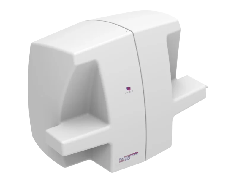
Key Features
- Dual brightfield and fluorescence scanning with a 12-slide capacity, ensuring efficient toxicology biomarker analysis.
- Automated workflows improve reproducibility and speed.
Pannoramic 1000 Digital Scanner
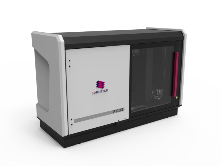
Key Features
- High-throughput scanning (1200 slides) ensures scalability for large toxicology studies.
- Z-stack and water immersion options allow for detailed cellular imaging in toxicity assessments.
№2. Tissue Microarray (TMA) for High-Throughput Toxicological Screening
TMA molmed

Key Features
- Automated tissue microarray creation supports batch analysis of toxicity effects on multiple samples.
- Direct molecular sampling allows researchers to extract DNA/RNA/proteins for further toxicological validation.
- High-density TMAs (up to 558 cores per recipient block) enable parallel analysis of multiple samples exposed to toxins.
№3. Advanced 3D Imaging for Structural Toxicology and Organ Analysis
Pannoramic-X

Key Features
- 3D virtual slicing & staining allows toxicity assessments without physical sectioning, preserving sample integrity.
- Biomarker quantification & volumetric analysis enable detailed organ-level toxicology studies.
- High-throughput scanning for full FFPE blocks or macro-blocks (up to 15 cm) enables large-scale toxicology research.
№4. AI-Powered Software for Data Analysis and Workflow Optimization
Software Solutions
SlideCenter & SlideMaster
Scalable, secure data handling for high-throughput toxicology research
Recent Case Study
Toxicology Study on Industrial Pollutants
Research Institution
National Environmental Toxicology Center
Objective
To assess the effects of industrial pollutants on liver tissue samples from animal models and human biopsy specimens.
Implementation
- Pannoramic 1000 Digital Scanner was used to scan 500+ liver tissue samples at 40x resolution for toxicology assessment.
- TMA molmed was employed to create high-density TMAs from affected tissues to enable batch biomarker analysis.
- Pannoramic-X Micro-CT provided 3D virtual staining and biomarker analysis, allowing researchers to study tissue damage without destructive sectioning.
- QuantCenter AI-powered analysis detected tissue fibrosis, inflammation, and necrosis levels, correlating pollutant exposure with histopathological alterations.
Results & Impact
- Improved Toxicity Assessment – Researchers achieved faster, more accurate analysis of pollutant-induced tissue damage.
- Higher Throughput & Reproducibility – Scanning and AI-based analysis reduced errors and increased efficiency by 60% compared to manual microscopy.
- Cost Savings & Scalability – Digital pathology reduced the need for physical slide storage and repeated analysis, saving 30% in lab costs.
- 3D Analysis Enhanced Understanding – Pannoramic-X’s 3D visualization enabled more detailed toxicology assessments than traditional histology.
The combination of 3DHISTECH’s digital pathology scanners, TMA solutions, 3D imaging technology, and AI-powered analysis software provides a game-changing approach to toxicology and environmental science research.
These solutions address key challenges such as throughput, accuracy, and scalability by enabling high-throughput toxicology screening, automated biomarker quantification, and AI-driven analysis. With the ability to handle thousands of toxicology samples with precision and efficiency, researchers, pharmaceutical companies, and regulatory bodies can make data-driven decisions faster, with higher confidence and reproducibility.
By integrating digital pathology solutions, toxicology and environmental science can move toward more precise, scalable, and efficient research workflows, accelerating discoveries in toxin exposure, drug safety, and environmental impact studies.
Who Benefits from These Solutions?
Toxicologists
Faster, high-resolution analysis of toxic effects in tissues, automated AI-driven quantification.
Environmental Scientists
Better assessment of pollutants’ impact on biological samples, high-throughput screening.
Pharmaceutical Companies
Reliable preclinical drug toxicity testing, improved biomarker quantification.
Regulatory Agencies (FDA, EPA, EMA)
Standardized, reproducible digital pathology data for environmental risk assessments.
Academic & Research Institutions
Scalable toxicology workflows with remote collaboration tools and AI-driven data handling.