• Skip to primary navigation
  • Skip to main content

The Digital Pathology Company

The Digital Pathology Company

  • Solutions
    • Solutions for Diagnostics
    • Solutions for Research
    • Solutions for Education
    • Solutions for Veterinary Pathology
    • Solutions for Geology
    • Scanning Service
  • Diagnostics
    • PANNORAMIC Pathology Management System
      • CaseManager
        • ClinicalViewer
      • DiagnosticApplications
    • PANNORAMIC Diagnostic Scanners
      • PANNORAMIC Flash DESK DX
      • PANNORAMIC 250 Flash III DX
      • PANNORAMIC 480 DX
      • PANNORAMIC 1000 DX
      • Legacy Scanners
    • Laboratory Information System
      • Track & Sign
      • CentralLogService Application (CLS) – Cybersecurity
    • Laboratory Components
      • MacroStation
      • SlideDriver
  • Research
    • PANNORAMIC Digital Slide Scanners
      • PANNORAMIC 1000
      • PANNORAMIC 250 Flash III
      • PANNORAMIC Confocal
      • PANNORAMIC SCAN II
      • PANNORAMIC MIDI II
      • PANNORAMIC DESK II DW
    • Tissue Microarrayers
      • TMA Grand Master
      • TMA Master II
      • TMA Applications
        • TMA Control
        • TMA Module
    • Digital Microscopes – Viewers
      • SlideViewer
      • iPad Viewer
      • InstantViewer
      • 3DView
    • Image Analysis – QuantCenter
    • Slide Management – SlideCenter
      • SlideMaster
    • ESchool
    • Software Downloads
  • Sales
    • Newsletter
    • Inquiry
    • Distributor Network
    • Become a Distributor
  • Support
    • Technical Support Form
    • Activity Report Form for Partners
    • Knowledge Base
      • Reference Institutes
      • Publications
        • Publications
        • TMA Grand Master publication list
        • TMA Master publication list
      • Case Studies
      • Whitepapers
      • Tutorials
    • Request Demo
  • News & Events
    • Events
    • News
  • About
    • Awards
    • Quality Management System
    • Funds
    • Funds (HU)
    • Contact Us
  • Careers
  • Partner Home
  • Account
  • Log Out
Home / News / 3DHISTECH Releases QuantCenter 2.3 with Unique Image Analysis Module

3DHISTECH Releases QuantCenter 2.3 with Unique Image Analysis Module

2021.06.17. by

We are proud to announce the release of version 2.3 of our QuantCenter digital image analysis platform.

QuantCenter is our multiple-module image analysis platform designed for whole-slide quantification in histopathology and molecular pathology. Thanks to its wide range of optional modules, QuantCenter provides researchers with maximum flexibility in quantitative image analysis, contributing to world-class research results. What’s more, QuantCenter’s modules can be linked for multi-level analysis.

Now we are proud to announce the release of QuantCenter version 2.3 with the following key new features:

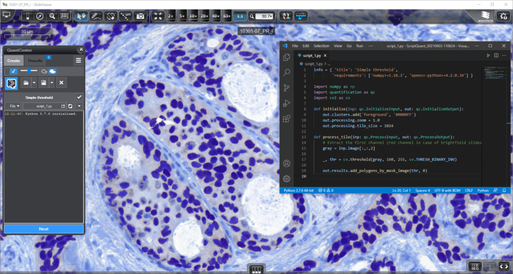
ScriptQuant: New image analysis module for maximum flexibility

ScriptQuant provides a shell for user-created image analysis scripts in Python for maximum flexibility for the research user, enabling them to run their own image analysis algorithms within the QuantCenter framework.

Its key features include

  • – Arbitrary script editor (VS Code by Microsoft is recommended)
  • – The user-created script can be saved into a scenario and handled like one
  • – Fast visual feedback: write your code and see the results immediately!
  • – ScriptQuant can be extended with external libraries (e.g. OpenCV, TensorFlow…)
  • – Well-documented API and use case examples, and many more

scriptquant

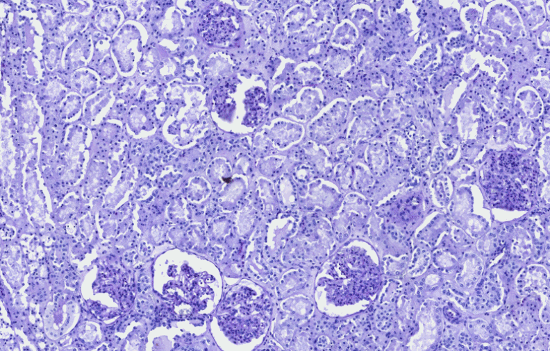
PatternQuant Plus: Image analysis for complex tissue segmentation projects

PatternQuant Plus is an improved alternative to the existing PatternQuant image analysis module – intended for use on more challenging samples (low visual difference between the staining of tissue types within the sample) and for complex tissue segmentation projects when thorough training using deep learning on multiple slides is needed. PatternQuant Plus can analyze both fluorescent and brightfield slides.

Its key features include

  • – Faster and more accurate tissue segmentation even in case of weak contrast and problematic staining
  • – Option for training on multiple slides
  • – Multiple classification features
patternquant plus
Image analysis of a low-contrast sample with PatternQuant Plus

For more information about the QuantCenter™ image analysis platform, please visit the QuantCenter product page. 

  • Scanning Service
  • PathoNet
  • Awards
  • Publications
  • Software Downloads
  • Newsletter
  • Contact Us
  • Careers
  • Sitemap
  • Privacy Policy
  • Terms of Use
  • facebook
  • twitter
  • linkedin
  • youtube
  • instagram

Partners

Login

© 2005-2023, 3DHISTECH Ltd. All rights reserved.
H-1141 Budapest, Öv u. 3., Hungary

We are using cookies to give you the best experience on our website.

You can find out more about which cookies we are using or switch them off in settings.

3DHISTECH Ltd.
Powered by  GDPR Cookie Compliance
Privacy Overview

This website uses cookies so that we can provide you with the best user experience possible. Cookie information is stored in your browser and performs functions such as recognising you when you return to our website and helping our team to understand which sections of the website you find most interesting and useful.

Strictly Necessary Cookies

Strictly Necessary Cookie should be enabled at all times so that we can save your preferences for cookie settings.

If you disable this cookie, we will not be able to save your preferences. This means that every time you visit this website you will need to enable or disable cookies again.

Google Analytics

This website uses Google Analytics to collect anonymous information such as the number of visitors to the site, and the most popular pages.

Keeping this cookie enabled helps us to improve our website.

Please enable Strictly Necessary Cookies first so that we can save your preferences!