The tissue microarray (TMA) technique is a valuable, high-throughput tool for diagnostic and research purposes. By being able to place up to several hundred different samples into one paraffin block, TMA saves time and costs of tissue preparation, slide preparation and staining.
With several TMA systems installed worldwide in pathology departments at hospitals, biobanks and research institutes, we are the leaders in the field of digital TMA.
Pathologists use the TMA technique as an excellent tool for cost effective diagnosis. TMAs are also extremely useful as internal controls (on-slide control) for traditional IHC slides.
Biobanks can create custom-made TMAs (tissue sample collections) for academic and pharmaceutical research as well. TMA blocks are widely used in pharmaceutical and biomedical research for the following applications: identification of predictive or prognostic factors, validation of newly discovered biomarkers, identification of diagnostic targets and therapeutic targets, antibody or probe validation.
TMA Hardware
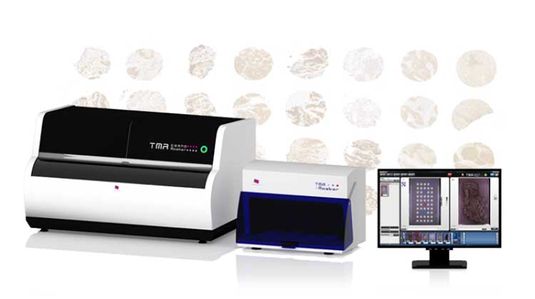
By producing smaller and higher capacity fully automated tissue microarrayers, 3DHISTECH is able to give a suitable TMA solution for the majority of customers.
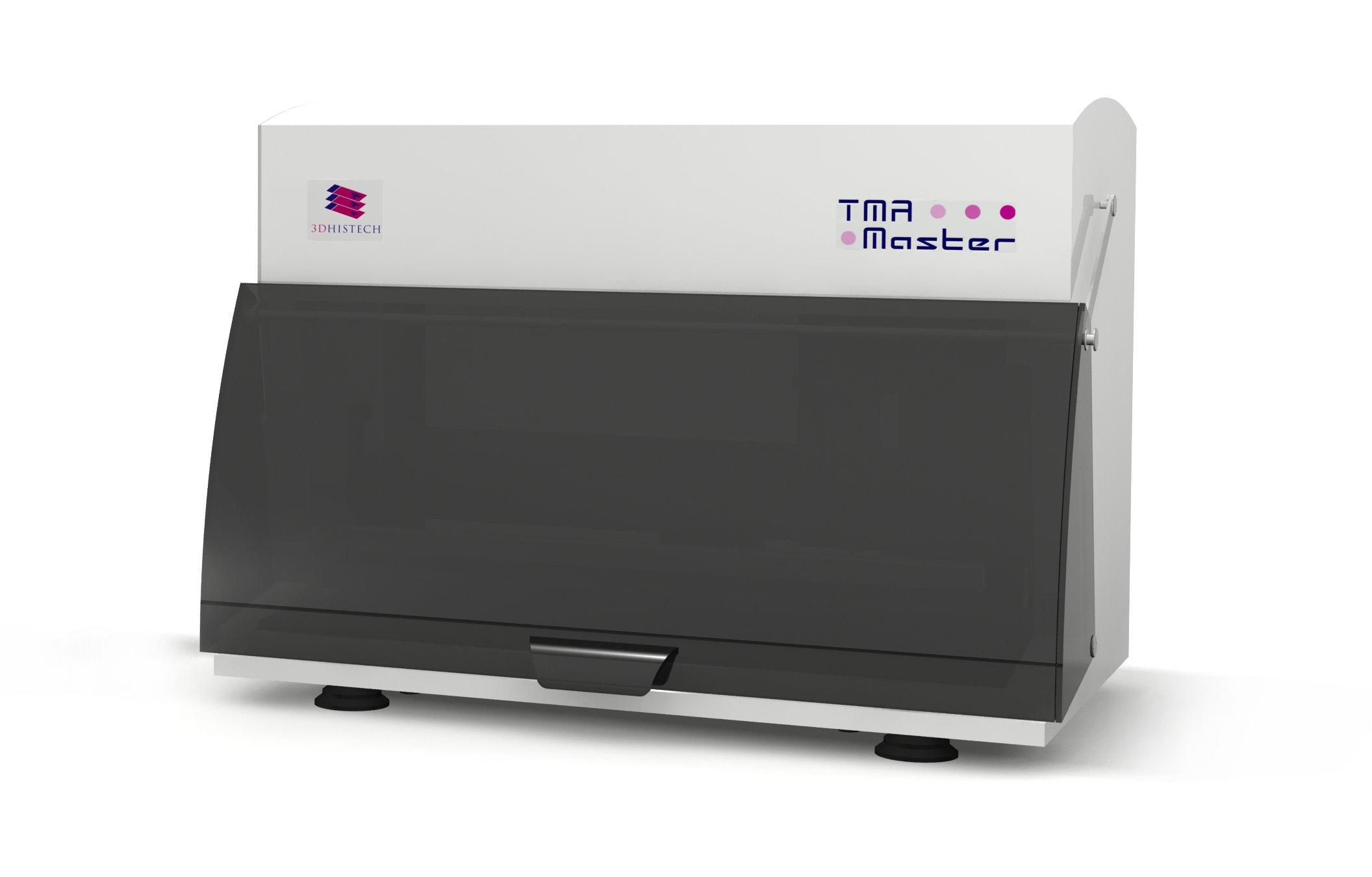
- TMA Master II is our smaller tissue microarrayer. It is the smallest fully automated tissue microarrayer on the market, it easily fits on any laboratory bench. With its 5 block capacity and high speed it is the ideal solution for smaller laboratories and pathology departments.
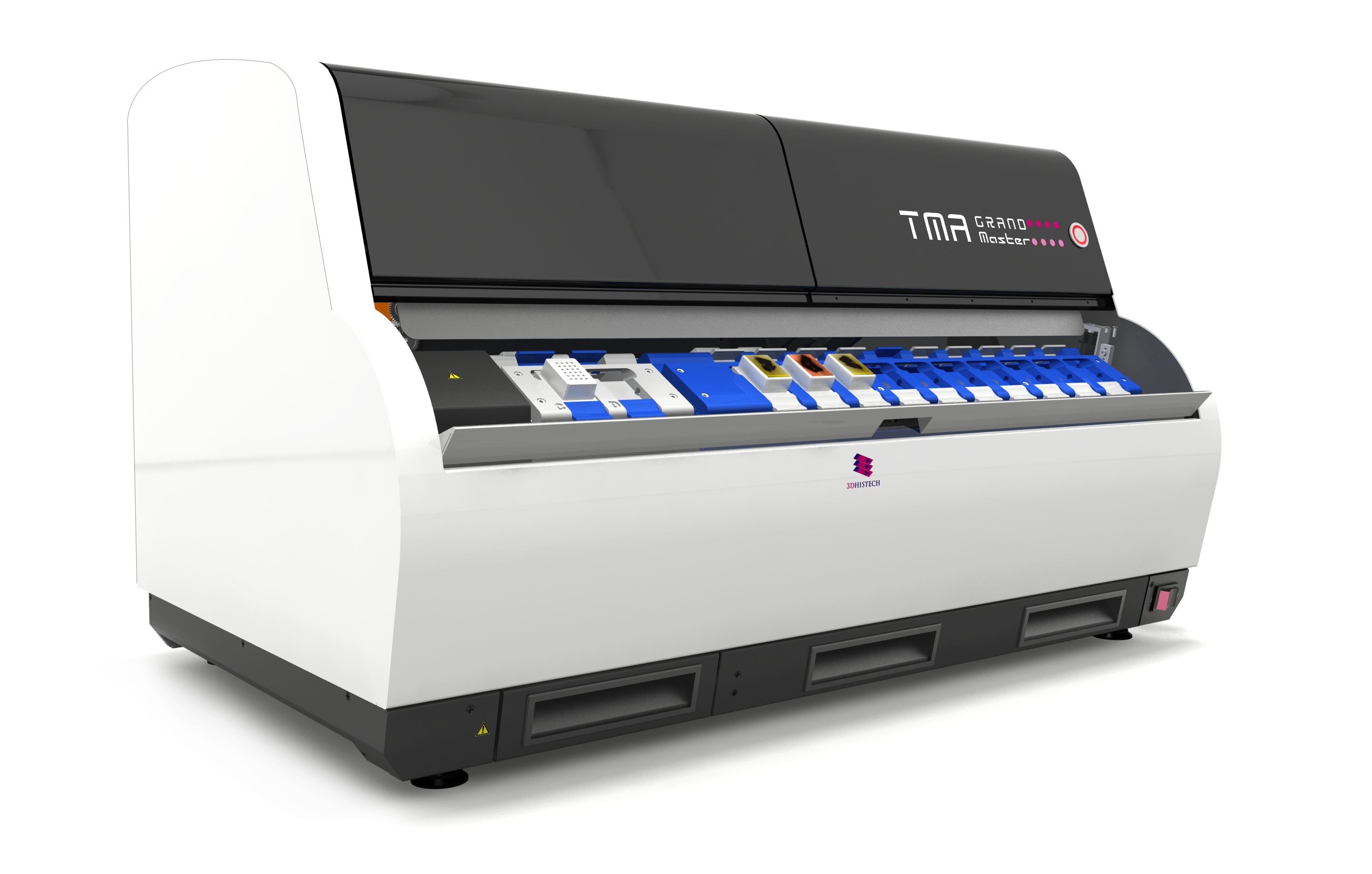
- TMA Grand Master is our flagship tissue microarrayer. Currently the TMA Grand Master is the highest capacity and the fastest tissue microarrayer at the market. TMA Grand Master is the ideal solution for biobanks, pharmaceutical companies and big pathology departments.
TMA Control software
The TMA Control software is the perfect solution for TMA block design and creation.
By using the TMA control software the operator can easily design the recipient block layout. Tissue core selection is also computer-assisted, the user can select the relevant tissue cores from the donor block image. If needed, the pathologist can overlap a previously annotated slide image with the donor block image by using the automated Digital Slide Overlay functionality. During the coring process the software automatically records which tissue core has been placed in which recipient block position. At the end of the workflow, this TMA data can be exported in Excel file format, and will be an invaluable resource of information during the TMA slide analysis.
The TMA control software works with TMA Grand Master and TMA Master tissue microarrayers.
Key Features
- Project-based workflow
- Recipient Block layout designer
- Ability to import Donor Block ID and additional sample data from Excel file
- Barcode-based donor block identification
- Automatic digital slide search from SlideCenter or local drive
- Automatic Digital Slide Overlay with TMA markers from Viewer
- Ability to place tissue cores in a clean PCR tube for later molecular analysis.
- Advanced and customizable export tool: export TMA data with Donor Block images
TMA Module software
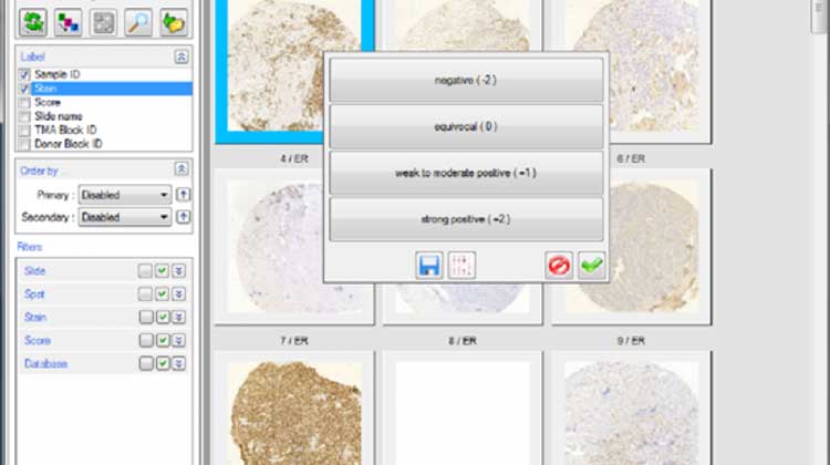
The TMA Module software is the ideal solution for TMA spot detection and analysis. The TMA Module can handle several hundred tissue cores. It is fully compatible with 3DHISTECH digital slide format and with the TMA database files created by our tissue microarrayers. During the spot detection the software is able to number and annotate the spots from the TMA database file, additional sample data is also overtaken and is available for later export.
The pathologist can set up various scoring schemes for manual scoring. A color can be assigned to each scoring level by the user. Based on the previously set scoring scheme the pathologist can score all the spots in a user friendly gallery window. After the scoring process, in the gallery window, each TMA spot will be marked with the previously set color according to the score selected by the pathologist. Then, the scored TMA spots can be filtered and ordered by various parameters such as: score, stain, sample ID, etc. After the scoring process, the scoring data, combined with the original TMA data, can be saved in Excel file format.
The TMA Module software automatically saves a project file, with all the spot finding and scoring data. Because of this the scoring process can be stopped any time and resumed later. By opening the TMA Module Project file the user can reopen and rescore any TMA slide.
By using the annotations generated by the TMA Module, QuantCenter applications can automatically quantify selected tissue cores. The results of the automated quantification will be saved directly to the digital slide and could be reviewed spot by spot or in total.
PCR Extraction
The PCR extraction functionality is one of the unique features of 3DHISTECH tissue microarrayers. By using this feature the pathologist is able to isolate and extract tissue samples in clean 0.2 ml PCR tubes. The extracted tissue samples can be used in the molecular pathology workflow. By using commercially available third-party kits, there is a possibility to perform DNA extraction or direct polymerase chain reaction (PCR) on these tissue samples. The DNA extracted from these samples can be used for any molecular biology application, such as quantitative polymerase chain reaction (qPCR), multiplex PCR and DNA sequencing.